The technology could help predict which tumours will light up on scans without the need for gadolinium, according to researchers.
Brain tumour patients could be spared contrast dye injections during MRI scans with the help of artificial intelligence (AI).
The technology could help predict which tumours will light up on scans without the need for gadolinium, according to researchers.
Gadolinium is a clear, colourless fluid injected into a patient’s bloodstream to make images clearer during an MRI scan and help with diagnosis.
However, the long-term effects of exposure on the body “remain unknown”, experts said.
There are also “broader environmental implications” linked to its use, they added, with gadolinium detected in sewage, surface and drinking water, water sources distant from MRI facilities, and fast-food soft drinks.
Experts at UCL developed an AI tool to predict exactly which parts of the brain would be enhanced if contrast fluid had been injected into the bloodstream using 11,089 MRI scans from more than 8,500 patients in the UK, US, the Netherlands and Nigeria.
On more than 1,100 non-contrast images, AI correctly predicted whether a tumour would brighten 83% of the time.
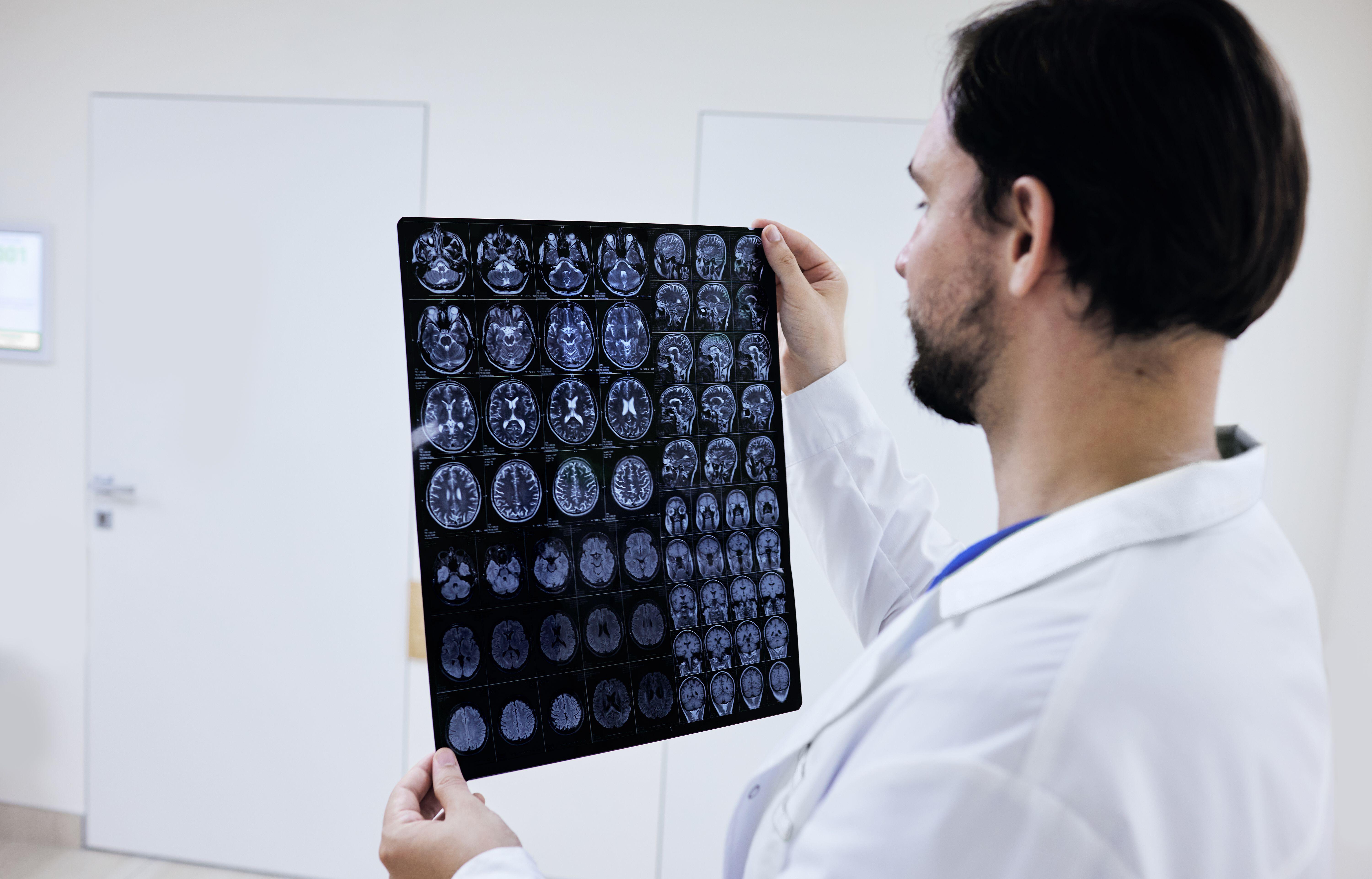
The study found the technology correctly identified 92% of tumours that did brighten and 74% that did not.
It was most accurate in scans of meningioma – a common brain tumour – but less accurate in cases involving children.
Researchers said: “These models show promise as a triage or decision-support aid – for example, flagging studies in which post-contrast enhancement is likely so that contrast can be added to a non-contrast-only protocol – and could reduce gadolinium dependence in neuro-oncology imaging.
“Future studies should optimise these models through close collaboration with expert radiologists.”
They added that the model’s performance is “currently insufficient to replace contrast-enhanced MRI” entirely, particularly in children, where is showed “reduced accuracy”.
However, it could be used as “a triage or decision-support aid” to flag where enhancement is likely, which would help “patients avoid a second hospital visit and minimise delays to patient care”, researchers suggest.
Dr Karen Noble, director of research and policy at Brain Tumour Research, said: “More than 100,000 people in the UK are estimated to be living with a brain tumour or the long-term impact of their diagnosis, with many of them undergoing regular MRI scans to monitor tumour growth.
“AI innovation that could reduce the need for contrast dye injections during MRI scans for some patients is welcome.
“We look forward to understanding how this model could be used in future to improve care, minimise side effects and help inform treatment decisions for brain tumour patients.”